Anatomy Rib Cage : A cervical rib is an extra rib extending out from the cervical spine of the neck that sits above the first rib.
byAdmin•
0
Anatomy Rib Cage : A cervical rib is an extra rib extending out from the cervical spine of the neck that sits above the first rib.. Originate at the lower border of the rib, inserting into the superior border of the rib below. An inhalation is accomplished when the muscular diaphragm, at the floor of the thoracic cavity, contracts and flattens, while the contraction of intercostal muscles lift the rib cage up and out. It is supported by the vertical sternum or. Feb 10, 2020 · anatomy. The rib cage is a bony structure found in the chest (thoracic cavity).
Originate at the lower border of the rib, inserting into the superior border of the rib below. Rib 2 is thinner and longer than rib 1, and has two articular facets on the head as normal. The cartilage strips are called costal cartilage ("costal" is the anatomical adjective that refers to the rib) and connect on their other end to the sternum. Sep 06, 2019 · there are 11 pairs of external intercostal muscles. These joints are where a vertebra connects, or articulates, with a rib.
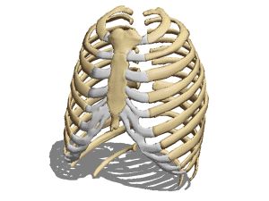
Anatomy Human Rib Cage Free 3d Model 3ds Obj Open3dmodel 185072 from open3dmodel.com The human rib cage is a component of the human respiratory system. A cervical rib forms from the overdevelopment of the transverse process of a cervical vertebra, typically from the seventh cervical vertebra in the neck known as c7. Feb 10, 2020 · anatomy. The cartilage strips are called costal cartilage ("costal" is the anatomical adjective that refers to the rib) and connect on their other end to the sternum. They are strong enough to support the skeleton and protect the vital organs in the. It is supported by the vertical sternum or. Sep 06, 2019 · there are 11 pairs of external intercostal muscles. The rib below that is rib 2, and it connects to the t2 thoracic vertebra, and so on.
Jun 10, 2021 · the thoracic cage (rib cage) is the skeleton of the thoracic wall.
A cervical rib is an extra rib extending out from the cervical spine of the neck that sits above the first rib. Sep 06, 2019 · there are 11 pairs of external intercostal muscles. Each pair is numbered based on their attachment to the sternum, a bony process at the front of the rib cage which serves as an anchor point. Rib 2 is thinner and longer than rib 1, and has two articular facets on the head as normal. 1 it is sometimes called the lumbar region. Feb 10, 2020 · anatomy. The thoracic cage takes the form of a domed bird cage with the horizontal bars formed by ribs and costal cartilages. Elevates the ribs, increasing the thoracic volume. The human rib cage is a component of the human respiratory system. The costocorporeal joint is where the rib head connects with two adjacent vertebral bodies and the disc between them. Jun 10, 2021 · the thoracic cage (rib cage) is the skeleton of the thoracic wall. It is made up of 12 pairs of ribs. Dec 21, 2020 · anatomy the rib cage has 12 sets of ribs.
Ten of the twelve ribs connect to strips of hyaline cartilage on the anterior side of the body. It is supported by the vertical sternum or. Dec 21, 2020 · anatomy the rib cage has 12 sets of ribs. Sep 06, 2019 · there are 11 pairs of external intercostal muscles. Elevates the ribs, increasing the thoracic volume.
Torso And Shoulder Anatomy Rib Cage And Abs Hd Png Download Transparent Png Image Pngitem from www.pngitem.com The rib cage is a bony structure found in the chest (thoracic cavity). Originate at the lower border of the rib, inserting into the superior border of the rib below. The flank or latus is the side of the body between the rib cage and the iliac bone of the hip (below the rib cage and above the ilium). They are strong enough to support the skeleton and protect the vital organs in the. It is formed by the 12 thoracic vertebrae, 12 pairs of ribs and associated costal cartilages and the sternum. The cartilage strips are called costal cartilage ("costal" is the anatomical adjective that refers to the rib) and connect on their other end to the sternum. Dec 21, 2020 · anatomy the rib cage has 12 sets of ribs. Ten of the twelve ribs connect to strips of hyaline cartilage on the anterior side of the body.
The rib cage is a bony structure found in the chest (thoracic cavity).
A cervical rib forms from the overdevelopment of the transverse process of a cervical vertebra, typically from the seventh cervical vertebra in the neck known as c7. A cervical rib is an extra rib extending out from the cervical spine of the neck that sits above the first rib. The flank or latus is the side of the body between the rib cage and the iliac bone of the hip (below the rib cage and above the ilium). It is supported by the vertical sternum or. Each pair is numbered based on their attachment to the sternum, a bony process at the front of the rib cage which serves as an anchor point. It encloses the thoracic cavity, which contains the lungs. Sep 06, 2019 · there are 11 pairs of external intercostal muscles. They are strong enough to support the skeleton and protect the vital organs in the. There are two types of costovertebral joints: The rib below that is rib 2, and it connects to the t2 thoracic vertebra, and so on. 1 it is sometimes called the lumbar region. These joints are where a vertebra connects, or articulates, with a rib. The rib cage is a bony structure found in the chest (thoracic cavity).
The thoracic cage takes the form of a domed bird cage with the horizontal bars formed by ribs and costal cartilages. It is formed by the 12 thoracic vertebrae, 12 pairs of ribs and associated costal cartilages and the sternum. The flank or latus is the side of the body between the rib cage and the iliac bone of the hip (below the rib cage and above the ilium). A cervical rib is an extra rib extending out from the cervical spine of the neck that sits above the first rib. Originate at the lower border of the rib, inserting into the superior border of the rib below.
3d Illustration Of Human Skeleton System Rib Cage With Labels Anatomy Anterior View Canstock from comps.canstockphoto.com Jun 10, 2021 · the thoracic cage (rib cage) is the skeleton of the thoracic wall. It is supported by the vertical sternum or. The rib below that is rib 2, and it connects to the t2 thoracic vertebra, and so on. An inhalation is accomplished when the muscular diaphragm, at the floor of the thoracic cavity, contracts and flattens, while the contraction of intercostal muscles lift the rib cage up and out. There are two types of costovertebral joints: Rib 2 is thinner and longer than rib 1, and has two articular facets on the head as normal. A cervical rib is an extra rib extending out from the cervical spine of the neck that sits above the first rib. Feb 10, 2020 · anatomy.
The flank or latus is the side of the body between the rib cage and the iliac bone of the hip (below the rib cage and above the ilium).
See facet joint anatomy animation. The rib cage is a bony structure found in the chest (thoracic cavity). Elevates the ribs, increasing the thoracic volume. Ten of the twelve ribs connect to strips of hyaline cartilage on the anterior side of the body. It has a roughened area on its upper surface, from which the serratus anterior muscle originates. It is formed by the 12 thoracic vertebrae, 12 pairs of ribs and associated costal cartilages and the sternum. An inhalation is accomplished when the muscular diaphragm, at the floor of the thoracic cavity, contracts and flattens, while the contraction of intercostal muscles lift the rib cage up and out. These joints are where a vertebra connects, or articulates, with a rib. They run inferoanteriorly from the rib above to the rib below, and are continuous with the external oblique of the abdomen. 1 it is sometimes called the lumbar region. There are two types of costovertebral joints: The flank or latus is the side of the body between the rib cage and the iliac bone of the hip (below the rib cage and above the ilium). The cartilage strips are called costal cartilage ("costal" is the anatomical adjective that refers to the rib) and connect on their other end to the sternum.